Cellular imaging

FV1000 confocal
Tumor cells cluster actine cytosqueleton in displayed magenta, dapi stained nucleus @Stephanie Dauvillier (CNRS), MULLER team
Spining Disk
Extract of time lapse of Human airway organoids (AOs) control (up) & infected with dTomato-expressing Mycobacterium abscessus (bottom). @MEUNIER team
LSM710 confocal
Lipid droplets in breast tumor cells. (lipids in green, Actine in red & nucleus in blue) ©Marie Rebeaud (CNRS), MULLER team
LSM710 confocal
Protein relocalisation after DNA double strand damage induced with biphoton laser. ©Phillipe Frit (CNRS), BRITTON team
Axio Imager M2 Zeiss
Grass blade autofluorescence Widefield and with Apotome module (7µm section) ©TRI: Eve Pitot (CNRS)
FV1000 Widefield
Grass blade autofluorescence Widefield & after deconvolution ©TRI: Eve Pitot (CNRS)
Previous slide
Next slide

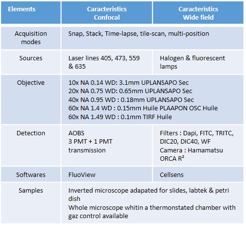
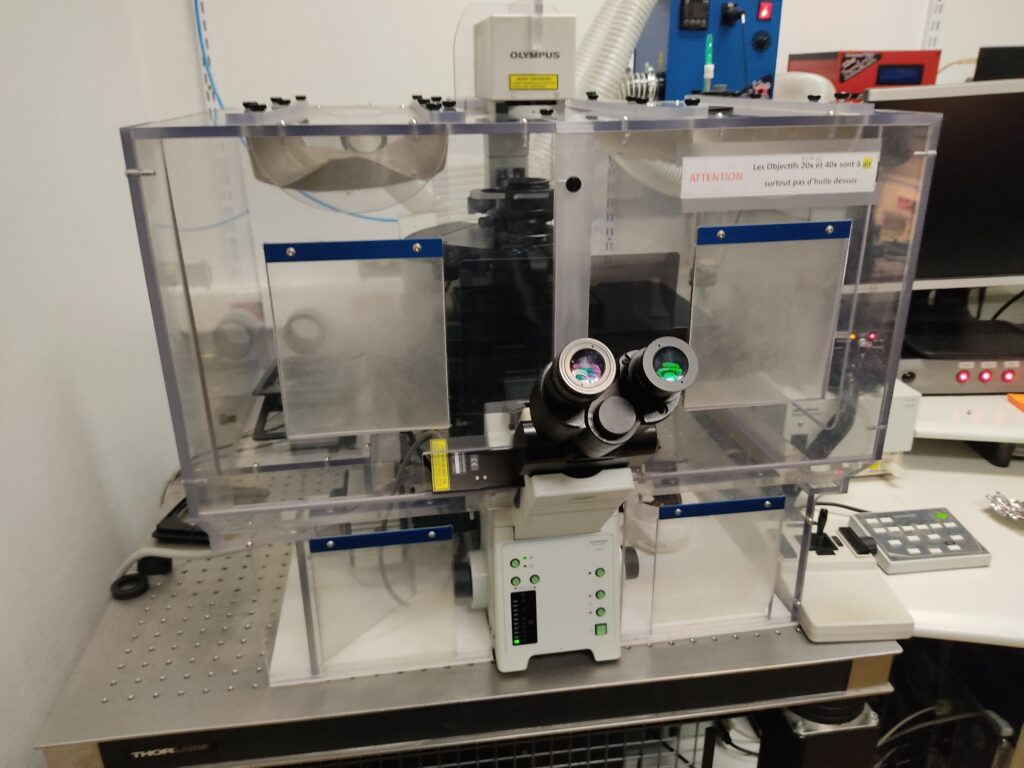
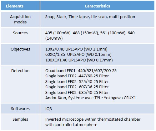
Cellular imaging allows high-resolution visualization of cellular and subcellular structures either in fixed or live specimens. For this purpose, our core facility offers wide field and confocal microscopy equipment to perform such type of analysis in a standard or BSL3 environment for the use of human pathogens.

In particular, we offer the possibility to perform FLIM and FRAP techniques in live specimens using a confocal laser scanning microscope equipped with femtosecond pulsed laser.

Confocal & Multiphoton Zeiss 710 NLO « Spectral »

710

Confocal & Widefield Olympus FV1000

Widefield & Apotome Axio Imager M2 Zeiss

apotome

Spinning disk Andor/Olympus in BSL3