

HIV-1/TB co-infection
Dissemination of HIV-1 in macrophages, perspectives in HIV/tuberculosis co-infection
A research priority for HIV eradication is the elimination of virus reservoirs. Macrophages are HIV-1 targeted cells involved in chronic activation of the immune system and establishment of persistent viral reservoirs, being a source of rebound of viremia after cessation of treatment. They are found in tissues as multinucleated giant cells (MGC), which are formed by fusion of infected macrophages [1]. We and others have also demonstrated that infected macrophages participate in the dissemination of the virus into host tissues. Indeed, we observed that HIV-1 dramatically alters all macrophage migration modes, inhibiting the amoeboid migration and enhancing the protease-dependent mesenchymal mode, effects under the control of the viral protein Nef [2]. In vivo, these observations translate into massive tissue infiltration of macrophages expressing Nef. Most of the work on HIV-1 infection of macrophages has been done with cell-free viral infection. However, cell-to-cell transmission is suspected to represent the dominant mode of infection in vivo. Importantly, this mode of virus transmission in macrophages may also allow the virus to escape antiretroviral drugs and the immune system. We recently investigated the cellular mechanisms involved in HIV-1 infection of macrophages and have shown that HIV-1 is efficiently transferred from infected T lymphocytes to myeloid cells through an actin-dependent adhesive structure, resulting in cell fusion and the formation of highly virus-producing MGC [3].
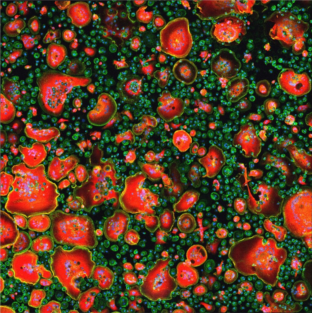
Co-infection with Mycobacterium tuberculosis (Mtb), the etiological agent of tuberculosis, and HIV-1 is major health issue in the world. The synergistic relationship between HIV-1 and Mtb is known to result in increased pathogen proliferation and associated pathogenesis, worsening diagnosis and treatment. We showed that HIV-1 infection is exacerbated in macrophages exposed to TB-associated microenvironments due to the formation of tunneling nanotubes (TNT) [4]. These cellular structures favor HIV-1 spread among macrophages by cell-to-cell fusion processes and again, formation of MGC. To identify molecular factors associated with TNT function and MGC formation, we performed a transcriptomic analysis in these macrophages, and revealed the up-regulation of type-I IFN signature [5], including the lectin receptor Siglec-1/CD169. We demonstrated the deleterious role of Siglec-1 in co-infection both in vitro and in co-infected non-human primates. Intriguingly, Siglec-1 expression localizes exclusively on a specific subset of long and stable TNT that contain HIV-1 cargo, and drives TNT-mediated HIV-1 transfer between macrophages [6]. Altogether, these mechanisms likely participate in enhanced virus load observed in HIV/TB co-infected patients, and Siglec-1 localization on TNT opens new avenues to better understand cell-to-cell viral spread and myeloid cell fusion.