

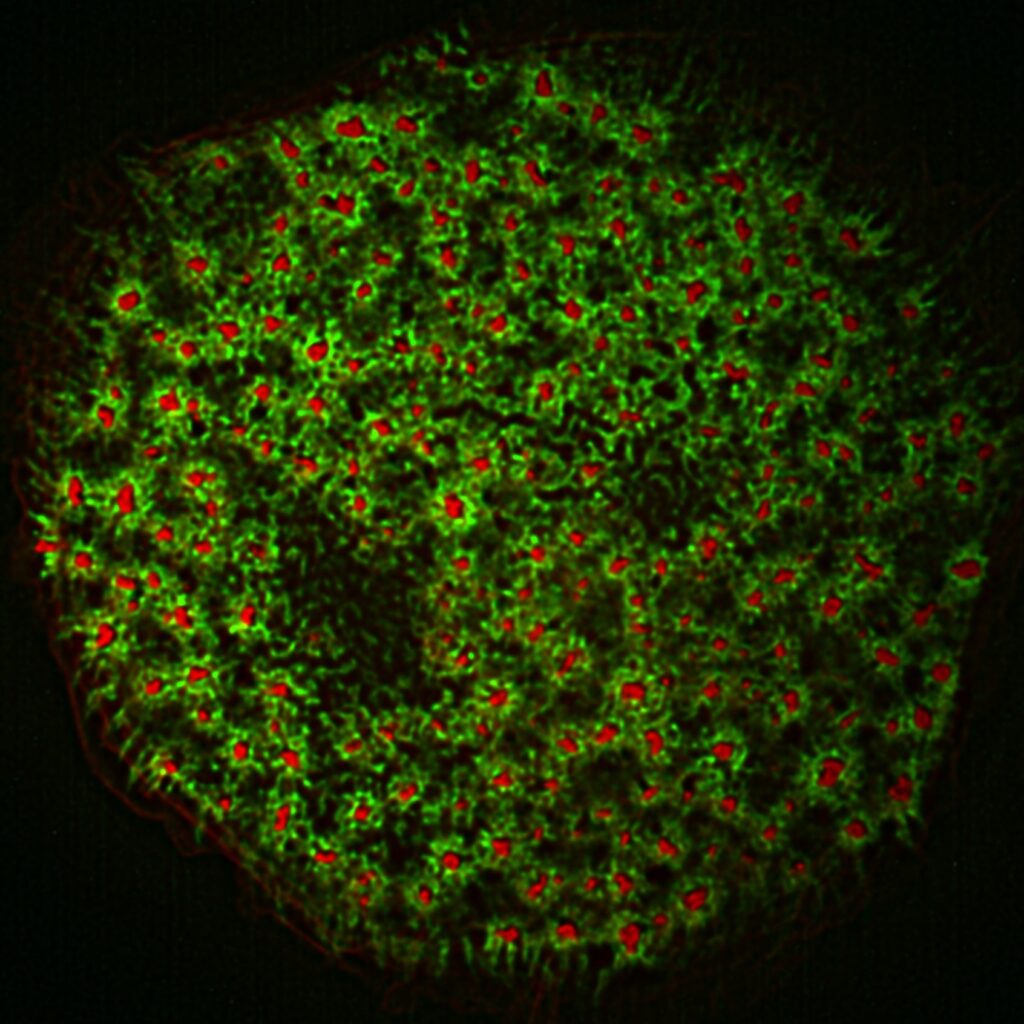
Podosome mechanics
In collaboration with C. Thibault and C. Vieu (LAAS CNRS) we have started investigating the mechanical properties of podosomes. Using atomic force microscopy combined with immunofluorescence we determined the stiffness of the podosome core and showed that it features periodic oscillations regulated by acto-myosin activity [1].
We then developed an original approach that consists in measuring, at the nanometer scale using atomic force microscopy, the deformations induced by human macrophage podosomes on an elastic film a few nanometers thick. Based on the measurements of the properties of this film and using a mechanical model adapted to the particular architecture of these adhesion structures, we were able to evaluate the amplitude of the protrusion forces implicated. Using this strategy, we could estimate, for the first time, the forces developed by individual podosomes and demonstrate that these forces are correlated to the film rigidity.
We observed that these forces are oscillatory and varies in a synchronous manner for podosome first neighbors, a result that correlates with phase synchrony of core F-actin temporal oscillations. This dynamic spatial coordination between podosomes suggests a short-range interaction that regulates their mechanical activity. We have also determined that both actin polymerization and myosin II-mediated contractility are major players in the production of force at podosomes, and proposed a theoretical model based on the equilibrium between these two force generators proposes an explanation of the podosome oscillatory behavior [2-4].
We then recently provided an experimental proof that such a force balance exists. We inhibited the expression of several podosome ring components and studied the impact on podosome protrusive ability using atomic force microscopy. This approach showed that adhesion ring integrity is crucial for protrusive force generation at the core of the podosome. The adhesion ring would thus operate as a handle that transmits to the environment the force produced by the protrusive core. Using a tridimensional nanoscopy technique called DONALD, we revealed that talin, one of the ring components, is vertically stretched within a molecular scaffold that connects adhesion receptors to the cytoskeleton and contains vinculin and paxillin. Talin stretching increases as the podosome generates higher protrusive forces, which proves that the ring is subjected to mechanical tension. This fundamental result offers a new perspective on the workings of this structure developed by macrophages to migrate through dense environments [5].
Finally, we used in situ cryo-electron tomography in collaboration with Marion Jasnin to unveil how the nanoscale architecture of macrophage podosomes enables basal membrane protrusion. Quantitative analysis of podosome organization demonstrated that the core is composed of a dense network of bent actin filaments storing elastic energy. Theoretical modelling of the network as a spring-loaded elastic material revealed that it exerts forces of a few tens of nanonewtons, in a range similar to that evaluated experimentally. Thus, taking into account not only the interface with the membrane but also the bulk of the network, is crucial to understand force generation by actin machineries. Our integrative approach sheds light on the elastic behavior of dense actin networks and opens new avenues to understand force production inside cells [6].