Single cell analysis of specialized blood vessels that mediate lymphocyte entry to lymphoid organs
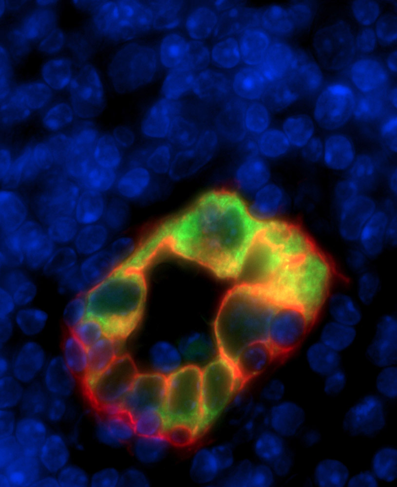
High endothelial venules (HEVs) are specialized blood vessels that play a critical role in immunity by allowing trafficking of lymphocytes through the different lymphoid organs of the body. HEVs are lined by endothelial cells with a plump almost cuboidal morphology. The team of Jean-Philippe Girard at the IPBS reports the first analysis of HEV endothelial cells by single-cell RNA sequencing and uncovers their molecular differences with endothelial cells from other blood vessels. This study was published in Cell Reports on March 12th 2019.
HEV endothelial cells are difficult to study because they are rare (less than 0.02% of the total cell population in adult mouse lymph nodes), and they rapidly lose their characteristics when isolated from their natural lymphoid tissue microenvironment (Moussion and Girard, Nature, 2011). In the present study, we overcame these difficulties by combining single-cell RNA sequencing, RNA-fluorescence in situ hybridization, and immunohistofluorescence. We demonstrate the cellular and spatial heterogeneity of HEVs at steady state, and we reveal that, after antigenic stimulation, HEVs undergo a global and temporary shift towards an inflammatory phenotype without compromising their ability to recruit naïve lymphocytes in inflamed lymph nodes. Most importantly we uncover differences in the regulation of genes critical for HEV-mediated lymphocyte trafficking. Blood vessels with HEV characteristics are found in solid tumors and chronically inflamed tissues (Girard et al. Nat Rev Immunol, 2012), and a better understanding of HEV differentiation and phenotypic diversity could have important implications for immunity, inflammation and cancer.
Reference
Single-cell analysis reveals heterogeneity of high endothelial venules and different regulation of genes controlling lymphocyte entry to lymph nodes. Veerman K, Tardiveau C, Martins F, Coudert J, and Girard JP. Cell Reports, 2019, 26:1–16 Read the paper
Contacts
Researcher l Jean-Philippe Girard l T 05 61 17 59 67 l Jean-Philippe.Girard@ipbs.fr
Press l Francoise Viala | T 06 01 26 52 59 | Communication@ipbs.fr