

Fight against tuberculosis: the structure of an important target unravelled
Despite its importance as a therapeutic target against tuberculosis, the structure of Pks13 (PolyKetide Synthase 13), an enzyme involved in the last steps of acid mycolic (MA) biosynthesis, was lacking. A study published in BMC Biology reveals the first structural information on the molecular mechanism of this complex enzyme and opens up new perspectives to develop inhibitors that target the interactions with its enzymatic partners or between catalytic domains within Pks13 itself.
The biosynthesis of MAs is an Achilles’ heel of Mycobacterium tuberculosis due to the essentiality of these components among Corynebacterineae species. Pks13 is the condensase required for the final condensation step of two long acyl chains leading to MA precursors. It is a megasynthase composed of several catalytic domains on the same polypeptidic chain, post translationally modified on the two acyl carrier protein (ACP) domains, leading to an holo-form. As well as other polyketide synthases, its structural study was challenging undoubtedly due to its inherent flexibility.
In order to decipher the Pks13 molecular mechanism, scientists used small-angle X-ray scattering of the entire protein and various molecular subspecies combined with known high-resolution structures of Pks13 domains or structural homologues. Taking into account the enzyme flexibility, these high-resolution structures have been located within the entire Pks13 enzyme. As a comparison, the low-resolution structures of two other mycobacterial polyketide synthases, Mas and PpsA from Mycobacterium bovis BCG, were also challenged.
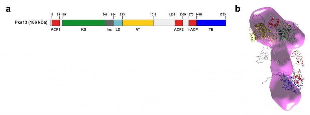
a. Domain organization of Pks13. Acyl carrier protein (ACP) domains: red; keto-synthase (KS): green; acyl-transferase (AT), yellow; thioesterase (TE), blue; hinge domain (LD), cyan; inter-domain hinges (gray).
b. Majority conformation of the M. tuberculosis Pks13 monomer. The atomic resolution structures obtained for the catalytic domains are superimposed on the full-size protein shell in pink. A flexibility hub of the enzyme is located at the interface between the ‘foot’ and ‘cap’ of the enzyme. © Bon & Mourey
This study highlights a monomeric and elongated state of the enzyme with the apo- and holo-forms being identical at the resolution probed. This may be required for the interaction with its known partners and/or to adjust the long acyl chains Pks13 has to condense. Nonetheless, the loading of a 16-carbon-long substrate analogue on one of its domain pushes the enzyme towards dimerization. Catalytic domains are segregated into two parts, which correspond to the condensation reaction per se and to the release of the product, a pivot for the enzyme flexibility being at the interface. Overall, the structural information may be critical to discover new drugs with novel mechanisms of action, targeting the interactions between Pks13 and its enzymatic partners or between catalytic domains within Pks13 itself.
Source
“Solution structure of the type I polyketide synthase Pks13 from Mycobacterium tuberculosis”
Cécile Bon, Stéphanie Cabantous, Sylviane Julien, Valérie Guillet, Christian Chalut, Julie Rima, Yoann Brison, Wladimir Malaga, Angelique Sanchez-Dafun, Sabine Gavalda , Annaïk Quémard, Julien Marcoux, Geoffrey S Waldo, Christophe Guilhot, Lionel Mourey
BMC Biol. 2022 Jun 21;20(1):147. doi: 10.1186/s12915-022-01337-9
Researchers’ contact
Cécile Bon | cecile.bon@ipbs.fr | +33 (0)5 61 17 58 40
Lionel Mourey | lionel.mourey@ipbs.fr | +33 (0)5 61 17 54 36
Press contact
IPBS : Françoise Viala | communication@ipbs.fr | +33 (0)6 01 26 52 59