

Molecule Delivery, Targeting and Diagnostic: new tools
Nanotheranostic tool for imaging and treatment of epithelial cancers
We are developing a nanocontainer protein as a vectorization system for the targeted delivery of therapeutic molecules and as a diagnostic imaging tool. The molecular marker specifically detected by this protein is characteristic of epithelial cancers. In collaboration with Toulouse Oncopole surgeons, we have chosen to provide proof of concept of this nano-tool efficacy on human ovarian adenocarcinoma. We validated the biochemical characterization of this nanocontainer. Four chemotherapeutic drugs were successfully confined inside the protein cavity and successfully delivered to adenocarcinoma cells in vitro. The use of this nanocontainer, as an imaging probe, after its labelling with a near infrared dye (Alexa 647), has been explored in vivo on a mouse model. We highlighted the possibility to detect submillimeter peritoneal xenografts of human tumor.
These results lead us to create the start-up See2cure in January 2024. See2cure was co-funded by G. Ferron (IUCT, CMO), L. Paquereau (IPBS, CSO) and M. Coustets (CEO). In December 2024, the Oncopole Board of Directors voted to invest in the capital of See2cure. One of See2cure’s objectives is to conduct a phase 1 clinical trial at the IUCT within the next 2 years.
Investigators: Former PhD M Coustets, Engineer & technician: C. Ladurantie; Researcher: M. Golzio, Project leader: L. Paquereau
Needle-free transdermal delivery
There are various alternative routes (electrical, mechanical, thermal) for transdermal delivery to avoid the use of the injection syringe, which could improve the quality of life of patients with diseases such as diabetes. These methods include, for example, micro-needles, electro-permeabilization, and iontophoresis. Electropermeabilization allows, via the application of an electric field, to temporarily increase the permeability of the skin and consequently to allow the transdermal passage of molecules of high molecular weight.
The goal of this ARN-funded project CARBO2DERM is to design and realize a nanocomposite patch based on carbon nanotubes (CNT) to store, but also releasing a drug when it is subjected to electrostimulation. To do this, different polymers and different shaping techniques have been explored and developed to demonstrate the feasibility of our approach.
Investigators: Former PhD student J Simon, Engineers: E. Bellard and G. Alberola; Researcher: M-P Rols and J. Kolosnjaj-Tabi; Project leader: M. Golzio
Collaborations : E Flahaut (CIRIMAT, Toulouse) ; Z Valdez (LAPLACE, Toulouse)
Optical imaging: multispectral, far-red fluorescent dyes, bioluminescence, intravital imaging and Speckle dynamic systems
Optical imaging of small animals allows observation of kinetic fluorescent events in time and space on the scale of a whole body, an organ or a cell. It enables detection and quantification to monitor gene expression, gene regulation, tumor growth and metastasis on a scale of days or weeks. We use and develop different approaches (multispectral imaging, fluorescent dyes emitting in the far red, bioluminescence).
The properties of the light can also be used. In collaboration with the ONERA, we develop a Tumor Microvasculoscope (TMV) based on Dynamic Polarised Speckle Acquisition (DYPSA). This process has been patented (Brevet 01/02/2017 n° FR 17 50833).
To overcome limitations induced by the skin, we also adopted the use of intravital microscopy with the implantation of a window chamber or dorsal skinfold chamber on the back of the mice. It allows the monitoring of fluorescent cell events in the skin or a cutaneous implanted tumor on several days in the same animal. Other window models exist that enable cells monitoring in deeper orthotopic tumors like abdominal window that we want to implement for in vivo electropermeabiliszation applications.
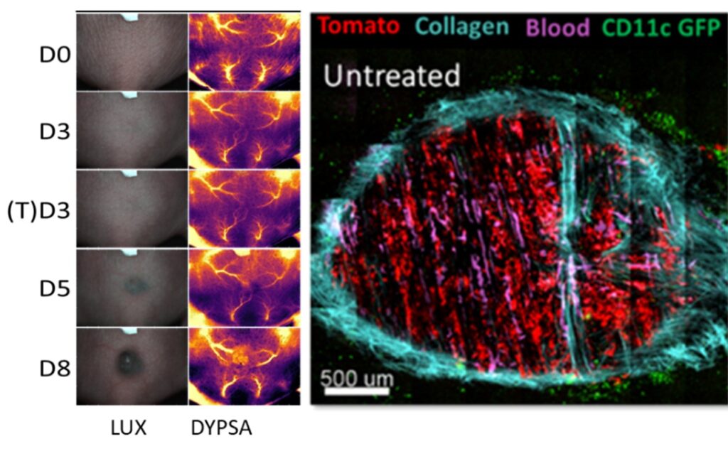
Left: Tumor photographs (LUX) and corresponding vasculature development images obtained via dynamic polarized speckle acquisition (DYPSA). Right: Micrograph showing a tumor in dorsal window chamber (DWC) observed with 2-photon microscopy. (unpublished data)
Investigators: Former engineer B. Jouanmiqueou, Former post doc N. Joncker, Engineer, E. Bellard; Project leader: M. Golzio
Collaborations: X. Orlik (ONERA, Toulouse)
Translation to the clinic
Early detection of cell death: We were able to provide elements of response through the early detection of cell mortality and therefore the effectiveness of electrochemotherapy (ECT) for cancer treatment (Calvel et al., IEEE 2024 and 2025) by microwave dielectric spectroscopy-MDS (Tamra et al., IEEE 2019) thanks to our work with the LAAS Toulouse (ANR Astrid RFMetrocell and RFMetrocell 3D projects). This innovative approach involved simultaneously applying EP electrical conditions to an individual cell while analyzing its cellular modifications in real-time using MDS and fluorescence microscopy within the same microdevice.
New protocols for cancer cell eradication: The collaboration with LEROY Biotech allowed us to develop and optimize new sequences of very high frequency pulses as effective as ECT (de Caro et al, J Control Release 2025), which no longer generate pain (de Caro, Bioelectrochemistry, 2023), and were compared to standard protocols of gene electrotransfer (de Caro et al, Pharmaceutics, 2023) in 3D spheroids models (Plan France Relance, SPEEDPORES).
Investigators: Former PhD students A Calvel, A de Caro, Engineers: E. Bellard and G. Alberola; Project leaders: M-P Rols and M. Golzio
Collaborations : D. Dubuc, K. Grenier (LAAS, Toulouse) ; J-B Leroy (LEROYBiotech, Saint-Orens de gameville)
Nanoparticle-based physical treatments to dismantle the extracellular matrix: from tumor’s microenvironment to bacterial biofilms
Cancer and infections are caused by malignant cells and the locally secreted extracellular components, into which therapeutic agents fail to penetrate. The impermeability of the tumor stroma is due to the dense extracellular matrix, particularly the collagen network. Conversely, bacteria form the biofilm, a dense macromolecules slime that prevents the penetration of antibiotics.
In order to dismantle the collagen matrix or biofilms and alleviate mechanical constraints to improve cancer or antibiotic therapy, this project presents an array of methods based on nanoparticle-mediated physical approaches, which could be used to treat highly desmoplastic tumors or bacterial infestations.
The objective of this project is to assess the efficacy of spatially and temporally controlled nanoparticle-based treatments exploiting the intrinsic physical properties of nanosized injectable (iron oxide) nanoparticles. These nanoparticles can generate heat when exposed to a laser (Kolosnjaj-Tabi et al., Cancers 2019) and magnetic field (Kralj et al., Adv Health Mater 2025) in order to exert physical stimuli- heat or mechanical forces, to disorganize the extracellular matrix. In a multifactorial approach, combining thermal, mechanical and electrical actions, this project aims to dismantle the tumor stroma or bacterial biofilms.
Proposed strategies are optimized and tested in vitro in two- and three-dimensional cellular models, with the goal of determining how altered extracellular matrix and, concomitantly, altered mechanics, could impact the outcome of prospective physical treatments. The efficacy of the treatment is monitored over time from the ultrastructural level to the macroscopic level.
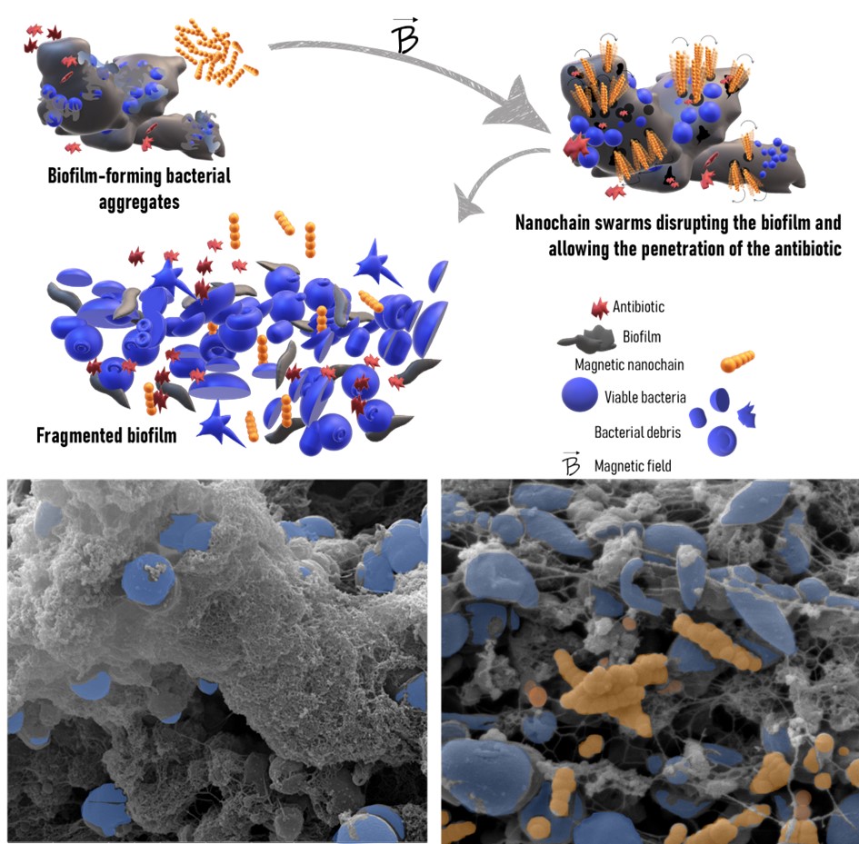
Schematic representation and scanning electron micrographs showing the collective action of magnetic nanochains to disrupt bacterial biofilms, adapted from Kralj et al., Adv Health Mater 2025.
Investigators: Former engineer C. Da Silva, PhD students E Barrère, N Mattei, Engineer: O. Cordier, Project leader, J. Kolosnjaj-Tabi
Collaborations: S. Kralj (University of Ljubljana, Slovenia), M. Chavent (CBI, Toulouse).