In the fields of cancer, infectious diseases and inflammatory diseases, our scientific objectives are based on two complementary lines of research: at the tissue and cellular levels, of the microenvironment in disease and its influence on treatment; at the molecular level, we study the molecular and structural mechanisms of disease, in order to characterize targets and propose candidates for new therapeutics.
Biology of the tissue and cellular microenvironment
This research axis covers the following topics:
Microenvironment in cancer, inflammation and infectious diseases: role in protection and susceptibility to disease, and impact on treatment,
Immune and inflammatory cells in cancer, inflammation and infectious diseases,
Eukaryotic and bacterial cell metabolism in cancer and infection.
Structural and molecular mechanisms of disease
This research axis covers the following topics:
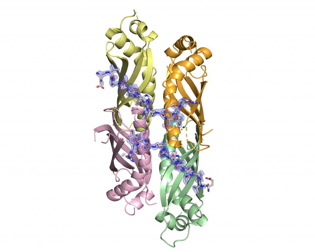
Structure-function-activity relationships of proteins and protein complexes in eukaryotic cells and bacteria, and rational design of functional ligands for the treatment of cancer and infections,
Genome stability,
Eukaryotic and bacterial lipids, membranes and envelopes,
Drug tolerance and resistance in cancer and infection.
Within the framework of these two research axes, methodological developments is pursued for the study of the microenvironment in cancer and infection, and for the structural and biophysical study of molecules and molecular complexes involved in cancer and infection.
Tissue engineering approaches such as organ-on-chip, organoids and 3D tissue printing that combine cells with engineered materials is used to study and manipulate organ functions and improve strategies for drug delivery by physical methods such as pulsed electric fields and radiofrequencies.
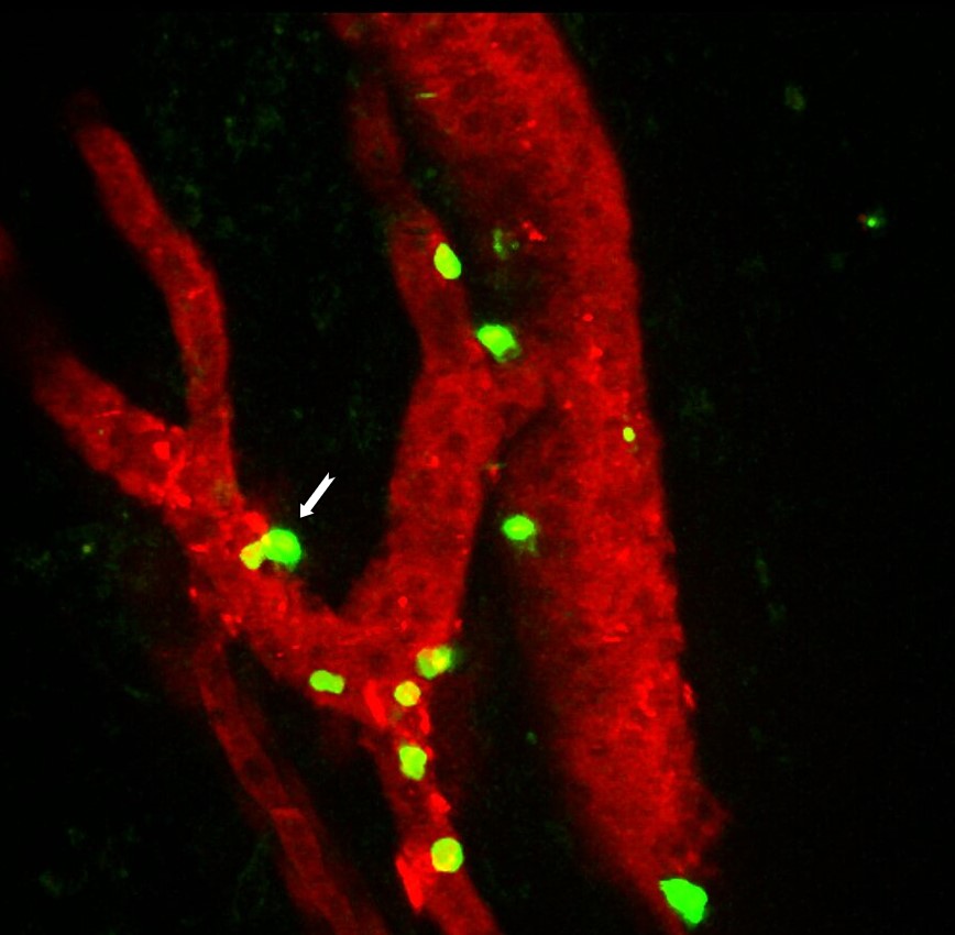
Altogether, the use and development of correlative imaging and tissue engineering technologies supported by the current expertise in optical microscopy, mass spectrometry and molecular imaging by FRET/FLIM techniques allows a better understanding of tissue and cellular microenvironments in tumours and infected tissues.